Hippocampus
Core of H.M.’s medial temporal resection story.
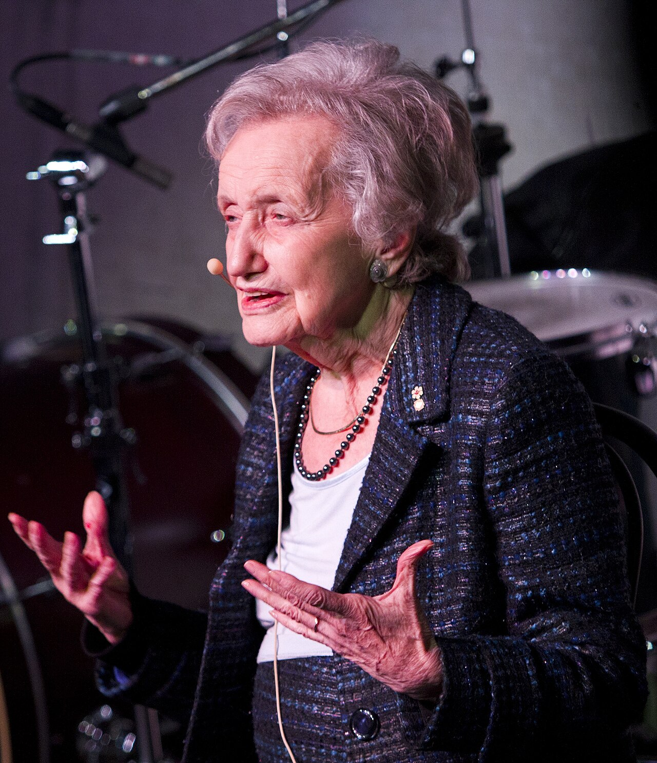
Brenda Milner · Cognitive neuroscience pioneer — patient H.M., memory systems and the lasting lesson that lesions can dissociate what looks unified.
Reviewed 2026-08·Sources & research·Methodology·Media credits
Darker cells mean a higher score for this topic on that metric.
LessMore
Last reviewed Sources & creditsMedia creditsMethodology
Henry Molaison (1953 bilateral medial temporal surgery). He lost new episodic memory while retaining skills — the dissociation that remade memory science.
1957. The paper tied hippocampal removal to anterograde amnesia without collapsing language or IQ in the teaching summary.
H.M. improved at mirror-tracing across days without remembering the sessions — procedural skill without episodic recall.
No. History of published cognitive neuroscience. Not medical advice, diagnosis, or surgical counsel.
Montreal Neurological Institute — careful testing, frontal-lobe work, and mentorship alongside the H.M. archive.
Hippocampus (memory index) and frontal lobes (control). Use dual-view; do not invent a personal lesion map.
The anatomical lesson of Milner’s era is medial temporal: hippocampus and neighbouring cortex for declarative encoding — not the whole brain as a single memory store.
Later imaging refined which subregions matter; the original dissociation still anchors teaching maps.
Core of H.M.’s medial temporal resection story.
Neighbour cortices also implicated in declarative memory.
Resected tissue included amygdala; affective lessons are nuanced.
Milner’s wider programme on strategy and memory control.
Systems supporting procedural gains H.M. could show.
Where consolidated remote memories can survive MTL loss.
Classic hippocampal loop for encoding.
Framework for why remote memories can remain.
Support procedural learning dissociated from episodes.
Other amnesia anatomies that refine the MTL story.
Historical surgical maps of H.M.’s lesion.
Iconic demonstration of intact skill learning.
Quantify anterograde declarative loss.
Later anatomy refined what was actually removed.
Open atlas neighbours: hippocampus, entorhinal, amygdala, frontal.
The neuron doctrine — drawing the brain cell by cell.
Research 58 🗣️Speech production and the birth of modern localization.
Research 69 👂Comprehension aphasia and the language network idea.
Research 58 🗺️Cortical maps from the operating room — the sensory homunculus.
Research 85 👁️How visual cortex builds edges, columns and critical periods.
Research 58 🐌Learning as synaptic change — from Aplysia to human memory.
Research 93 ❤️Feeling, self and somatic markers in decision circuits.
Research 78 🎨Vision, art and how cortex separates colour, form and motion.
Research 74 🧪Nerve growth factor — discovering how neurons survive and wire.
Research 97 🗺️Place cells and the cognitive map inside the hippocampus.
Research 51